About
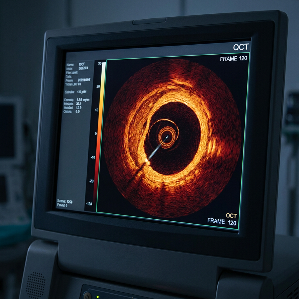
Coronary OCT Hub curates real‑world coronary artery disease cases, combining image atlases, stepwise interpretations, and teaching slides to make advanced intravascular imaging accessible for interventional cardiologists, fellows, and cath lab teams worldwide.
About
Coronary OCT Hub curates real‑world coronary artery disease cases, combining image atlases, stepwise interpretations, and teaching slides to make advanced intravascular imaging accessible for interventional cardiologists, fellows, and cath lab teams worldwide.
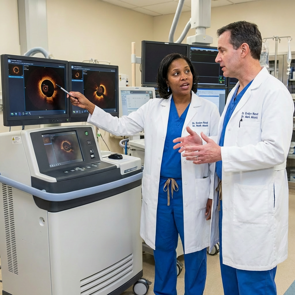
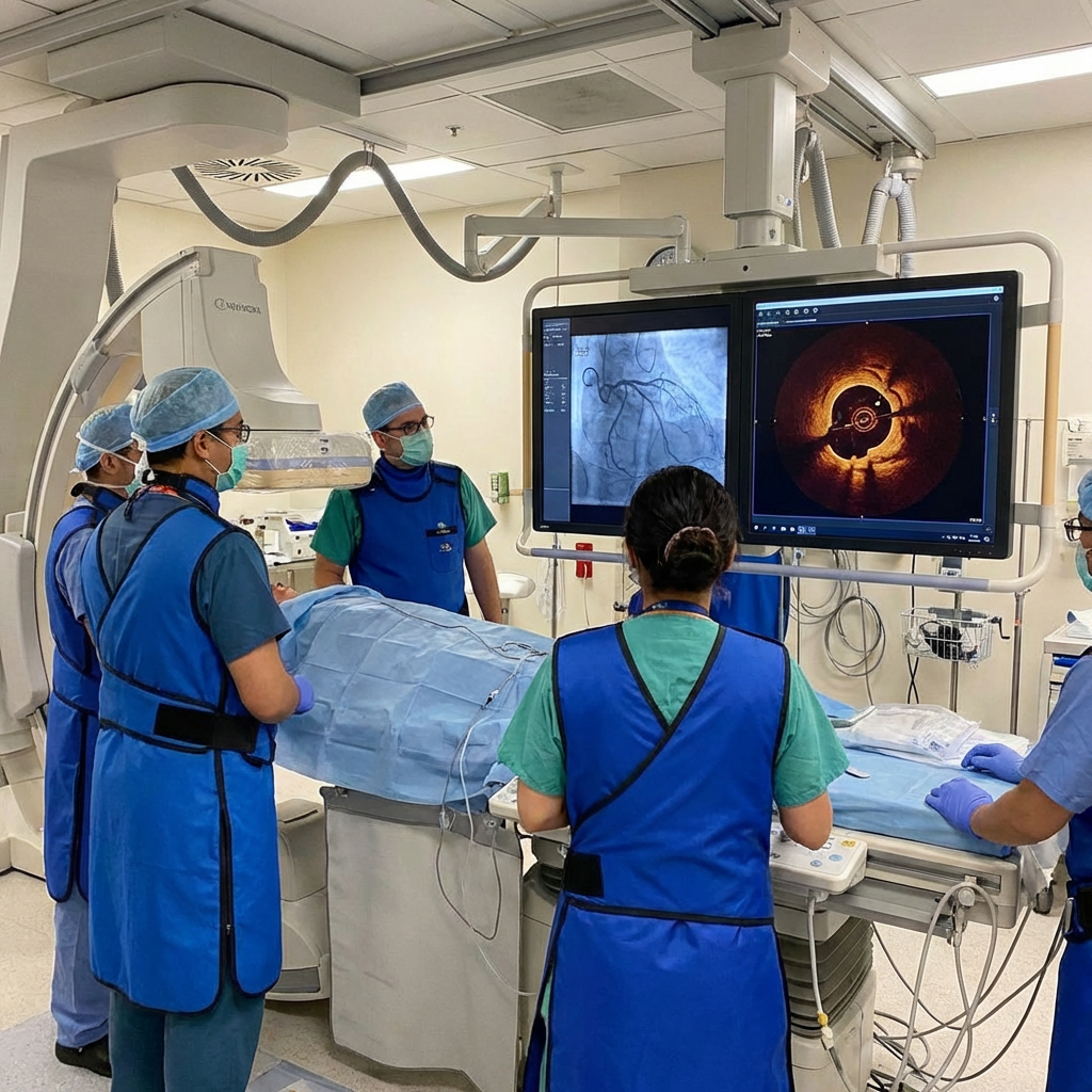
Medical professionals collaborate to analyze detailed cardiac imaging results in a clinical setting.
Cases
Resources
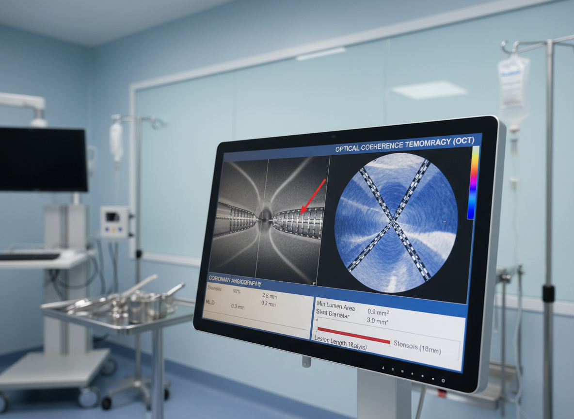
Interactive OCT case library with lesion assessment, stent optimization examples, and key teaching points for review before or after procedures.
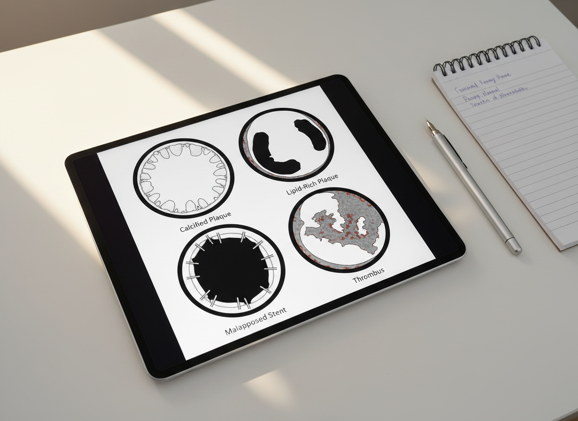
Downloadable OCT teaching slides, including classic patterns, pitfalls, and step‑by‑step interpretation frameworks for conferences, journal clubs, and trainee education.


